White line lesion is the second most common claw disease affecting dairy cattle, with 5.5 cases per 100 cows per year recorded as the average treatment rate in the UK1.
The average case is thought to cost approximately £193, costing the average farm over £1000 per year per 100 cows². However, in some herds white line can become a major problem with over 30 cases per 100 cows per year costing approximately £5800 per 100 cows per year.

Fig 1: A typical white line lesion in the heel of an outer hind claw. Note the stone has probably got stuck in the white line after the separation formed, but is now making the lameness worse
Causes
The white line is the softer, off-white horn that joins the wall horn with the sole horn. It is produced by the laminae which means any disturbance to the laminae, such as bruising can lead to a weakened white line. As the white line joins the hard capsule of wall horn with the relatively flexible sole horn, it has to withstand considerable physical tensions. These tensions and brusings are increased with turning and pushing behaviour on hard surfaces, especially if there are or sharp stones in the environment. Wet conditions can soften the horn and increase the risks as can thin soles through excessive wear and over-trimming.
The main theories thought to contribute to white line lesions include:
-
Any factors leading to poor horn quality which may be nutritional or environmental.
-
Physical trauma producing bruised white line horn
-
Shearing forces across the white line, such as twisting and turning on an unyielding surface
-
Loose stone and uneven surfaces that physically penetrate the white line
When the white line defect tracks as deep as the quick, an infection can establish, often resulting in an abscess if the drainage is blocked. Alternatively, a lower grade focus of inflammation or infection may become established present, producing a milder lameness.
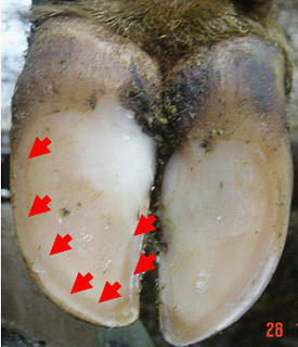
Fig 2: a picture with arrows showing the white line, the off-white horn that connects the sole with the wall horn.

Fig 3: the white line horn is deposited as the wall hornslides down 5 mm per month from the coronary band to the sole over folds of laminae, The main weakness often lies where thee is infilling between each of the laminae. This is visible as railway track markings on the white line where dirt penetrates the weaknesses. These weaknesses allow the white line to split running right up into the laminae in lameness cases. A seat of infection can then establish high up the wall, as seen in this claw.
At least 80% of white line lameness cases occur in the outer claw of the hind feet in most feet, mostly in the heel and outer side of the claw, reflecting increased exposure to exposure to physical forces in this claw at this site.
Treatment
Dutch 5 step method foot trimming should be performed, followed by careful examination will help identify the site of pain and the white line lesion. Animals exhibiting signs of moderate to severe lameness could have an abscess.
Fig 4 (above and below) Many white line abscesses form at the base of very fine tracts in the white line. Treatment involves removal of the wall horn to create a drainage hole for the pus. Sometimes the track is hard (or impossible) to find.

Sometimes the place where infection enters may be hard to find. Heat and pain in the heel should help identify the affected claw. Hoof testers and thumb pressure around the wall and coronary band can identify the most likely white like track to follow.
Once the likely white line lesion causing lameness is identified, it should be opened by removing the wall from the sole surfaceup. This requires a very sharp disinfected knife, starting with the wall at the sole surface, being careful not to cut into the quick (see figures below). If there is an abscess, this will lead to the sudden release of pus. A large drainage hole should be made and weight relieved onto the opposite claw with a block. While it may be argued that bearing weight will help express pus, what happens in practice is that the cow avoids bearing weight and slurry blocks the drainage hole. A block on the healthy partner claw is always recommended. As Treponemes causing digital dermatitis can infect these lesions, antibacterial treatments should be applied regularly on exposed quick. A bandage and poultice for one day can help draw pus for severe lesions but this is not essential. The quick will be painful but also swollen, which can block the drainage hole, so anti-inflammatory treatment should be given. If significant improvement is not seen within 24-48 hours then the track will need to be re-explored to create better drainage. Good hygiene at trimming is essentail with clean disinfectant used on equipment and flushed on lesions. Cows that have had an abscess invariably benefit from further trimming of loose horn within 4 weeks to prevent wall ulcers. Very few require antibiotics unless infection is tracking deeply (indicated by swelling above the claw).
1-4 weeks following a white line abscess the extent of the under-run horn can be seen. The quick will be less swollen, and more easily avoided with the knife. The loose horn can be completely removed to avoid a chronic infection becoming established.

Fig 5a & b After 1-4 weeks days the swelling of the quick will have reduced allowing any residual under-run horn to be completely removed reudcing the risk of a wall ulcer. If this under-run horn is left, then slurry and moisture will be trapped next to the quick, encouraging chronic infection and creating the ideal environment for Treponemes (see Digial dermatitis module).
When white line abscesses have been missed, they either burst out the coronary band or run under the sole and burst out the back of the heel. If that is the case, then in most instances all the under-run horn has to be removed. When a deep and chronic infection of the quick in the wall has become established (wall ulcer) then veterinary advice should be requested as these are best treated under local anaesthesia by careful removal of dead tissue and infected leaves of horn (lamellae) (Figure 6c).

Fig 6a-c: In some cases, white line infections may track up the wall (left image) or under the sole to the heel (centre image). If the quick becomes chronically infected with Treponemes then any delays to treatment reduces the chances of full recovery partly due to the damage to the bones. The latter are very painful and require treatment using local anaesthesia. In all cases a block should be applied to the sound claw under -run horn needs removing and the quick should be cleaned to promote recovery.
To summarise, the principles of white line lesion treatment involve:
- Careful identification of the painful lesion
- Good drainage of any pus
- Relieve weight from the painful claw (apply block)
- Removal of any loose or under-run horn (in some cases1-2 weeks later)
Prevention of white line lesions
Prevention of white line lesions involves reducing factors leading to bruising with better lying, cow flow and walkways (tracks) and improving claw quality.
-
Reducing standing times - the longer cows stand on concrete, whether at milking, due to heat or due to problems with cubicles, the increased risk of bruising and risk brusing on concrete.
-
Cow flow - pushing, twisting and turning forces on the claw are likely to bruise and damage the white line. Common problems and solutions include:
-
Pushing on tracks - allowing cows to drift in without being hereded, gentle herding methods, wider tracks, more comfortable and cow-friendly tracks.
-
Pushing in collecting yard - improve collecting yard design to ensure good flow; entice rather than drive cows into the parlour, never shout or push cows; disarm the electric backing gate, train cows to move to a bell/buyzzer, only using the backing gate to take up slack space; avoid any aversive experiences.
-
Turning on the parlour exit - widen or straighten to improve dispersal and cover concrete with rubber matting.
-
Bullying in the housing - create cross passages, open up blind ending alleys, create extra space to reducing stocking density, address sources of bullying. (Figure 8)
-
-
Under-foot surfaces - improvements can usually be seen with gentle herdin g which allows cows to pick their routes to avoid stones. (See Cow Track module).
-
Diet - Dietary biotin supplementation at 20 mg per head per day has been shown to reduce lameness due to white line by up to 50% (3) and may also incrtease milk yield(4) partly or fully off-setting the cost. Supplementation needs to be maintained long-term and the improved claw health will only occur after approximately 130 days. Some people have speculated that subacuteruminal acidosis may remove natural syunthesis of biotin, as biotin synthesis may be increased with feeding hay. However, this is still unproven. The other important dietary intervetion is to minimise body condition score loss, which may alter the digital cushion increasing bruising and all claw horn lesions.
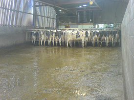
Fig 7: A misaligned heifer against a backing gate can be a risk for white line injuries. On this farm gentle use of the backing gate and rubber floor matting reduces the negative impact of this.
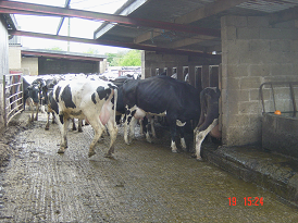
Fig 8: Out-of-parlour feeders are common places for cow bullying with twisting and turning.
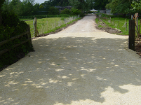
Fig 9: - Above is a picture of an oolitic limestone track. This material crushes to a fine material and self-stabilises. This can be achieved with hard stones and builders rubble (metal removed) by finely crushing, adding cement and compacting. R|eclaimed astroturf can be used to address abrasiveness. There are many locally available options and so for further information see the Cow Track module

References
1 Barker
2 Wilshire and Bell
3 Thomas and others
4 Nottingham digital cushion work - 3 papers
