Traumatic Reticulitis (wire, hardware disease)
Traumatic reticulitis occurs sporadically in adult cattle following ingestion of sharp metal and plastic objects (eg. fence wire and nails) and their localisation in the reticulum.
Outbreaks of disease have been reported after incorporation of wire from disintegrating car tyres used on silage clamps into the mixer wagon, and after access to bonfire sites.

Outbreaks of traumatic reticulitis have been reported after incorporation of wire from disintegrating car tyres used on silage clamps into the mixer wagon.
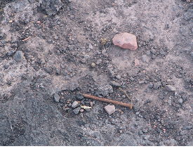
Nail left after bonfire - cows find the bonfire ash surprisingly palatable and often lick at such sites.
Clinical presentation
Classical clinical signs are only observed when the foreign body is in contact with the peritoneal lining of the abdominal cavity and may last only 2-3 days after which adhesions restrict reticular movement. There is sudden and complete loss of appetite, and a dramatic fall in daily milk production e.g. from 40 litres to 2-3 litres. The animal stands with an arched back and moves reluctantly, and is typically last to enter the milking parlour. The cow shows evidence of anterior abdominal pain with a taut rigid abdomen, refusal to turn sharp corners, ears back, and a fixed glazed stare. The cow is constipated and defaecation and urination are often accompanied by grunt. A pain response can usually be elicited when the cow's back is dipped behind the withers.

There is sudden and complete loss of appetite, and a dramatic fall in daily milk production e.g. from 40 litres to 2-3 litres.

The animal stands with an arched back and moves reluctantly.
Differential diagnoses
Your veterinary surgeon will carefully consider:
-
Peritonitis
-
Liver abscessation
-
Endocarditis
-
Chronic suppurative pneumonia
-
Pleural inflammation/abscess
Diagnosis
A diagnosis is based upon clinical findings and confirmed during surgery. Peritoneal fluid sampling and ultrasonography may be used to identify any peritoneal exudate/fibrinous reaction caused by the penetrating foreign body.
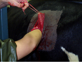
The diagnosis is confirmed during surgery.
Metal detectors are a waste of time due to the presence of harmless pieces of metal in the reticulum (anthelmintic boluses for example) and some objects may be plastic or other synthetic, non-metallic material.
Treatment
A course of parenteral antibiotics is very unlikely to be effective without surgical removal of the penetrating foreign body. Magnets administered orally will collect loose metallic objects in the reticulum but will probably not draw out objects firmly embedded within the reticular wall.
Surgery
Prompt surgery is essential to avoid the consequences that may result in culling for poor production or death due to septic pericarditis should the wire penetrate the sac surrounding the heart.
The piece of wire/nail/plastic can be recovered during surgery undertaken by your veterinary surgeon if the cow is presented soon after ingestion - delays can prove fatal.

Large piece of wire in the wall of the reticulum.
Recovery of milk yield is often slow due to the localised peritonitis present prior to surgery interfering with reticular contractility and propulsion of digesta. It may take the cow up to four weeks to regain her previous milk yield. The cow is treated with parenteral antibiotics (penicillin) for at least five consecutive days.

Neglected traumatic reticulitis case - this cow had extensive peritonitis when presented for veterinary investigation (see necropsy findings below).

Extensive peritonitis following neglected case of traumatic reticulitis.

Large accumulation of pus in the sac (pericardium) surrounding the heart following penetration by piece of wire from the reticulum.
Prevention/control measures
Magnets can be given to all cows by mouth which lodge in the reticulum and are reported to be highly effective in attracting ingested metal objects.
Septic pericarditis
Septic pericarditis occurs sporadically in adult cattle following ingestion of sharp, usually metal objects that migrate through the reticular wall (see above) and diaphragm into the pericardial sac (surrounding the heart).

Sharp wire penetrating the heart wall having migrated from the reticulum.

Large accumulation of pus in the sac (pericardium) surrounding the heart revealed at necrospy.
Clinical presentation
Affected cattle present with a history of reduced appetite, weight loss and reduced milk yield of several days' to weeks' duration. There may have been no clinical signs of traumatic reticulitis observed by the farmer. The cow is dull and depressed, and walks slowly. There are accumulations of subcutaneous fluid under the brisket and mandible (bottle jaw).

Large accumulations of subcutaneous fluid under the brisket and mandible.
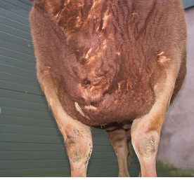
Considerable brisket oedema in this cow with septic pericarditis.
Differential diagnoses
Your veterinary surgeon will carefully consider:
-
Endocarditis
-
Myocarditis/dilated cardiomyopathy
-
Lymphosarcoma involving the mediastinum and pericardium
-
Thymic lymphosarcoma
Diagnosis
Pericarditis is diagnosed on clinical examination, and sometimes using ultrasound.
Treatment
Antibiotic therapy will not resolve the pericardial infection although temporary reduction of oedema occurs after a single corticosteroid injection such as dexamethasone and antibiotic therapy.

Antibiotic therapy will not resolve the pericardial infection.
Management/Prevention/Control measures
Take great care with building work - nails are a common foreign body. Prevent access to bonfires. Take care not to put tyres from the silage clamp into the forage wagon. Routine control measures relate to prevention of traumatic reticulitis where magnets are given per os to lodge in the reticulum and collect metal debris.
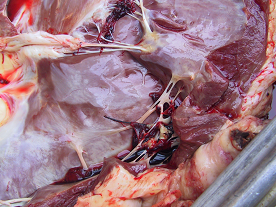
Sharp wire penetrating the heart wall having migrated from the reticulum.
Vegetative endocarditis
Infection of the heart valves proves difficult to diagnose in cattle in many cases.
Animals typically present with poor milk yield for several days to weeks and weight loss. Affected animals appear slow and stiff with elbow abduction, and have a poor appetite.
The provisional diagnosis of vegetative endocarditis is based upon clinical findings of chronic weight loss, pyrexia, and multiple joint effusions unresponsive to antibiotic therapy although temporary improvement lasting three to five days may be achieved after dexamethasone injection. Necropsy reveals cardiac enlargement and ventricular hypertrophy/dilation. The vegetative lesions are readily demonstrable on the heart valves.
Treatment
Treatment of vegetative endocarditis cases is invariably unsuccessful with the diagnosis confirmed at necropsy.
Management/Prevention/Control measures
Prevention of endocarditis is based upon timely and effective treatment of focal bacterial infections remote from the heart but these may, in themselves, not present with outward clinical signs e.g. metritis, mastitis, foot abscess, and grain overload.
Dilated (Holstein) cardiomyopathy (DCM)
Dilated cardiomyopthy is occasionally reported in well-grown 2-3 year-old Holstein cattle. Occurrence in progeny of certain bulls indicates a genetic component.
Clinical presentation
Clinical signs include marked peripheral oedema, jugular distension, and fluid accumulations in the body cavities.
Diagnosis
Necropsy reveals enlargement of the heart with a rounded "globose" shape. There may be no histological changes.
Treatment
There is no treatment for DCM. Occurrence in progeny of certain bulls indicates a genetic component


