Introduction
Maedi Visna (MV) is classed as one of the “iceberg diseases” of sheep which are found throughout the UK that are relatively infrequently reported but cause serious economic losses without ever being identified. In general, the onset of disease is slow with vague clinical signs; flock efficiency may be significantly affected but is often overlooked because of the lack of overt disease. The levels of MV in the UK is relatively unknown, recently studies have estimated 2.8% of flocks are affected, although within infected flocks up to 85% of animals may test positive for the virus. The disease is infectious and incurable.
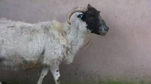
Maedi Visna may not present with specific clinical signs apart from chronic weight loss in older ewes
What causes MV?
Maedi Visna is caused by a small ruminant lentivirus, similar to Caprine arthritis encephalitis virus (CAEV), which has an incubation period of months to years. There are two distinct clinical forms of the disease: the respiratory form (maedi) and the neurological form (visna). Both types of disease are caused by the same virus. In the UK the respiratory form is most common, with older ewes (over 3 years of age) showing slow-developing pneumonia and weight loss. In addition to the main manifestations of MV, affected sheep can also suffer from mastitis and joint infections.
Infected animals mount an antibody response to the virus but are never able to eliminate it, so become lifelong carriers of the disease, spreading it mainly via respiratory secretions and milk. The virus can also be found in saliva, semen and urine, but it is unlikely these contribute significantly to the spread of infection.
Economic and Welfare impacts
There have been many studies which show the impact of MV infection on the overall flock production. Conception rates, milk production, lamb birthweight, and the weaning weight of lambs have all been shown to be reduced in flocks with active MV infection. Lamb mortality rates are also higher in these flocks. It is estimated that ewes are culled up to a year sooner than they would have been if not infected with MV, and the value of those cull ewes can be reduced by as much as 20% due to chronic wasting and poor condition.
Whilst mortality of animals with MV is low as a direct result of the infection, infected animals are more susceptible to secondary infections such as bacterial pneumonia. Similarly, factors such as husbandry and nutrition affect the overall mortality in a flock with MV.
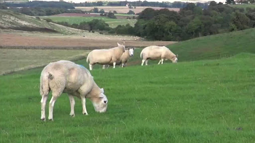
Affected animals are thin despite good nutrition
Clinical signs and diagnosis
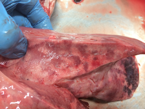
The respiratory form of the disease (maedi) is characterised by a chronic progressive pneumonia in older sheep, and associated weight loss and wasting. The pneumonia gets worse until affected sheep have difficulty breathing and eventually die, often with concurrent infections.
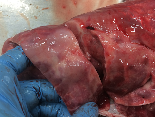
Typical lung lesions are often identified at post mortem, alongside secondary bacterial infections. Image credit: Ben Strugnell
In the neurological form (visna), the infected sheep develop progressive neurological deficits (toe dragging and single limb paralysis, ataxia (wobbling/incoordination), and in severe cases full paralysis and recumbency. Other signs include fine head tremor, circling, progressive head tilt, visual impairment, and separation from the rest of the flock. Whilst the respiratory form of the disease results in prolonged (months-years) deterioration, the neurological signs usually progress faster, resulting in the euthanasia or death of the animal within weeks of the first signs becoming apparent.
Neurological signs are not seen commonly in the UK but are most frequently identified in flocks with high levels of infection and active cases of respiratory disease.
Other forms of MV such as mastitis and arthritis are much less common and do not seem to result in the same chronic wasting as the respiratory and neurological forms, although they may lead to premature culling due to poor performance.
Diagnosis is made either on blood samples, milk samples or post-mortem examination. Blood testing is the cheapest and easiest way to test live animals for infection, however there can be a significant time lag between initial infection and the production of antibodies which are detectable with the testing methods available. Similar tests can be carried out on milk, which may prove easier or cheaper in milking flocks.
Post-mortem examination of sheep that have either died or been euthanased, will reveal classic lung pathology (respiratory disease), which can be further tested to confirm changes to the cells, or the virus can be isolated from brain tissue if the animal in question was showing neurological signs.
In the UK there is a Maedi Visna accreditation scheme which aims to identify MV-free flocks by periodic screening of a proportion of the flock.
Closer examination of lung tissue under the microscope will confirm the diagnosis. Image credit: Ben Strugnell
Treatment
There is no treatment for MV; supportive treatment may be appropriate for concurrent infections, but the affected animal will never be “cured”, and as such any therapy is ultimately pointless.
Prevention
It is thought that infection is most commonly spread either by respiratory secretions such as mucus, or via milk, so lambs born to ewes infected with MV are highly likely to become infected early in life. It should also follow that flocks that are intensively managed or housed would be more likely to see the disease spreading rapidly, however although this scenario is quite often the case, there are also reports of MV spreading rapidly through extensively managed flocks which are never housed; close contact between infected and non-infected animals is only part of the picture, and more research is needed.

Early identification of carrier animals allows for strategic culling or separate management to reduce the spread of infection.
Identification of animals infected with MV, and their removal from the flock, along with any offspring is the only way to work towards total freedom from the disease. Occasionally it may be possible to prevent lambs becoming infected soon after birth by removing them from the ewe immediately after they are born and rearing them artificially. However this approach is not without its problems, and artificially-reared lambs often do not do as well, and are more susceptible to infections arising from unhygienic feeding equipment or inadequate nutrition.
Farms where MV is not present should maintain very strict biosecurity measures to avoid inadvertently buying infected animals on to the farm. Similarly, those farms with separate MV-accredited and non-accredited flocks must take extra precautions as infection can easily spread between the two groups. It is reported that MV-accredited flocks are twice as likely to have a breakdown if there are non-accredited sheep on the same holding than those accredited flocks with no other sheep on the farm.
All MV-accredited flocks have to abide by the rules of accreditation, including blood sampling of any bought-in animals within 12 months (preferably whilst still in quarantine), and it is recommended that they test any show animals at the beginning and end of each show season.
If high levels of infection are found within a flock, sometimes the only option is to cull the whole flock and re-stock (with MV-accredited animals). Regular screening and careful consideration of incoming stock, coupled with strict biosecurity measures significantly reduce the risk of MV entering the flock in the first place. Prompt investigation of higher-than-normal levels of pneumonia, mastitis, barren ewes, or chronic wasting, will also help to identify infection within the flock sooner rather than later, and thus reduce the economic losses associated with Maedi Visna infection.