Many of the diseases that cause problems on the rearing field continue into the release pen period. Additional comment is made on these when necessary.
Ascarid worms
Described in detail in part 4 - the rearing field.
Aspergillosis
Described in detail in part 4 - the rearing field.
Cause:
An infection of the respiratory tract caused by the Aspergillus family of fungi. In the release pen a potent source of this can be damp straw or wood chips left lying in the pen.

Fig 1. Woodchips in release pen represent a risk of Aspergillosis
Clinical signs:
Gasping. Stretching of neck. Death.
Treatment:
Nothing specific.
Prevention:
Do not use straw in release pens. Do not leave heaps of wood chip in the pen after high pruning or felling trees.
Ataxia - Pheasant Ataxia
Cause:
Believed to be viral, but not accurately identified.
Clinical signs:
Usually presents in poults 8 weeks or more. Affected birds show imbalance, walk backwards. Sometimes unable to stand. When fallen tend to spread wings out in an apparent attempt to stabilise themselves.
Treatment:
None
Prevention:
None
Avian Influenza
Described in detail in part 4 - the rearing field.
Avian Tuberculosis
Cause:
Infection by the bacterium Mycobacterium avium. Most commonly seen in mature birds.
A chronic, slow spreading bacterial infection, characterized by the formation of tumor like lesions, called granulomas or tubercles, in the organs. Found in temperate climates all over the world. Most birds, including poultry, game birds, songbirds, crows etc can be affected.
Pheasants for some reason are extremely susceptible.
Clinical signs:
Are usually only seen in birds over a year old, due to the slow progressive nature of the disease. Usually only a few birds will show clinical signs.
The symptoms depend on which organ systems are affected by the granulomas. Most commonly, a progressive weight loss is seen despite a good appetite, with a persistent diarrhoea and soiling of tail feathers. Eventually the birds will become emaciated and die.

Fig 2. Avian Tuberbulosis. The white nodules may be seen all over the abdominal contents. The liver lies above the spleen.
Infected birds excrete the organism in their droppings. Other birds can get infected by ingesting feed, water, litter or soil contaminated by these droppings.
Spread:
Can be brought in by wild birds, contaminated shoes or equipment, pig manure spread on fields, effluent from dairies or meat processing plants, or by rodents entering game pens.
Treatment:
M. avium is relatively resistant to antibiotics plus a number of disinfectants. Outside the body, it can survive for many years in soil, but it is killed by direct sunlight. Within a carcass, it can only survive for a few weeks.
Control:
Environmental conditions greatly affect the susceptibility of birds to TB. An inadequate diet, crowded, wet and unhygienic circumstances will predispose.
Infected birds must be culled, as treatment is ineffective. The carcasses must be incinerated. . Further use of the site should be avoided for at least 2 years.
Good rodent control will help.
Humans are considered to be highly resistant to Mycobacterium avium, but infection can occur.
Blackhead (Histomoniasis)
Histomoniasis is caused by a single cell organism called Histomonas meleagridis. Also commonly called "Blackhead" because of the disease in turkeys, where the soft parts of the head become discoloured. Partridges are more at risk than pheasants. Both adult and young birds can be affected.
Young birds are often found dead in good condition with little or no specific signs. In older birds the disease process is more chronic. Affected birds are lethargic, rapidly loose weight and become unable to fly. Some birds will have yellow diarrhoea. Mortality may reach 100%.
Post mortem:
The caecum is thickened, inflamed and often filled with debris (caecal cores).

Fig 3. Blackhead. Large bowel lesions
Peritonitis is often found. In later stages the liver becomes affected and necrotic circles are visible on the liver surface.

Fig 4. Blackhead. Liver lesions "Typical" white, slightly raised lesions in the liver
The organism dies very quickly after the bird dies, so it is very important to present fresh samples, or even better, sick birds showing specific symptoms, to get an accurate diagnosis.
Direct infection, by ingesting infected droppings or scavenging dead birds, is not very common as the organism is very fragile in the environment. Histomonas can live inside the eggs of the caecal worm, Heterakis, for many years. They are also able live in the muscle of earthworms that have eaten Heterakis eggs. Ingesting either these eggs or earthworms may infect birds.

Fig 5. Blackhead. Bundles of Heterakis worms in the blind gut
Treatment and control:
Has become difficult since the banning of Emtryl. Some clinical response has been reported following the use of oregano extracts (Herban®). This has been used in conjunction with tetracycline antibiotics.
Prevention is much more important, through good management and sanitation. Control of Heterakis should be carried out by routine worming, and proper cleansing and disinfection of pens and materials, wire mesh platforms for feeders and drinkers, etc.
Blepharitis - Tick mediated
Cause:
Bacterial infection following penetration of the skin by tick bites.
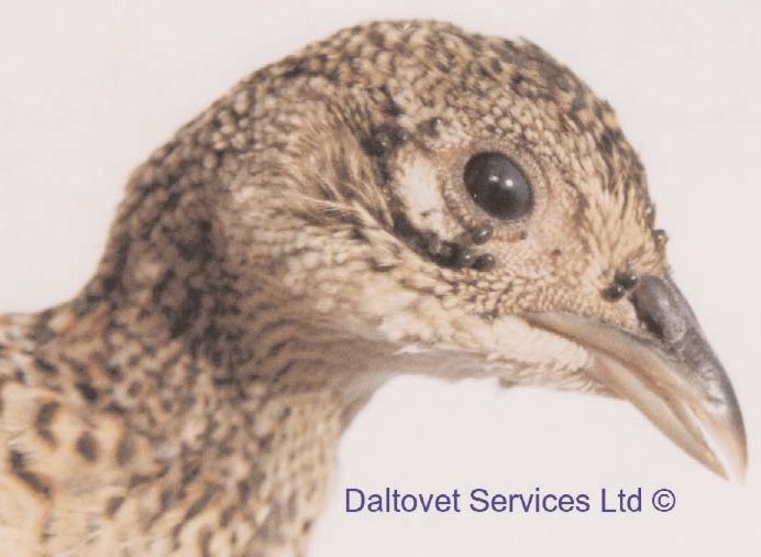
6a Numerous ticks around the eye
Clinical signs:
Swelling around the eyes. Can lead to complete closure and effective blindness. Birds can't feed and waste away.
Treatment:
Affected birds can be treated by application of topical insecticides originally designed for use in domestic pets. "Rubbing bars" of hessian sacking of similar material can be strategically placed near drinkers of feeders to treat the birds in situ.
Prevention:
Reduce tick population. Cut bracken and don't allow grass to become too long. Ticks attach to the host by grabbing hold whilst it brushes against the grass.
Beware ticks carry Lyme disease, which is a serious problem in humans. Avoid ticks bites. Pay close attention to marks persisting more than 48 hours. If you are bitten consult your doctor.
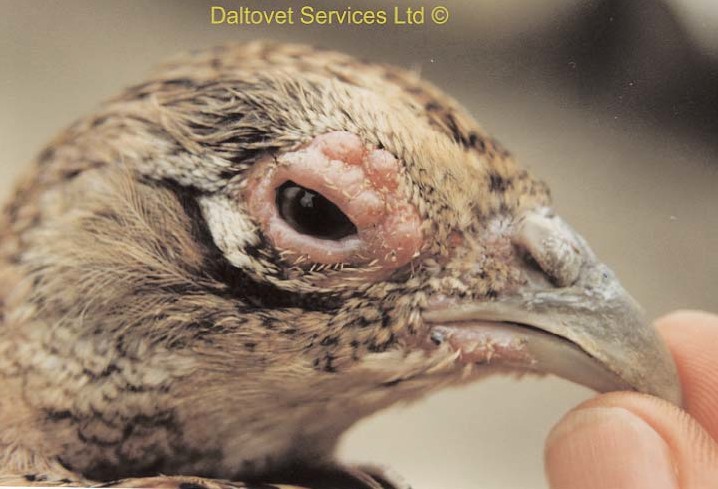
Fig 6b. Peri-orbital dermatitis caused by ticks.
Botulism
Cause:
Poisoning by toxins produced by Clostridium botulinum. This grows readily on dead meat and can also be found in the maggots following fly strike on dead bodies.
Clinical signs:
Early signs are of birds sitting down showing reluctance to move. Paralysis of the wings and legs follows, as well as the classical sign of Limberneck.

Fig 7. Botulism "Limberneck" Flaccid paralysis of the neck muscles
The eyelids and in particular the third eyelid are also paralysed meaning that there is no blink reflex when the cornea is stimulated. Take care when carrying this out, as it is painful and, of course the bird can show no evasive action. Mortality rates can be remarkably high.

Fig 8. Botulism. Third eyelid paralysis. A light touch to the cornea causes no blink
Treatment:
Success or failure depends mainly on how much toxin has been taken in. Mildly affected birds may recover provided they are isolated and supplied with food and water. Antibiotic and vitamin support may be of some help.
Prevention:
Don't allow dead birds to lie around and decompose (Fig 8A). Attempts at fly control are unlikely to be much help, so don't let them lay their eggs and so expose the birds to affected maggots.
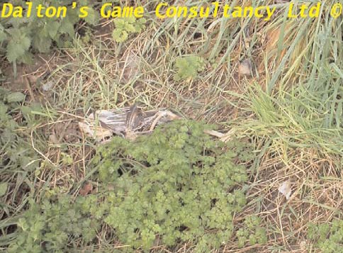
Fig 8a. Dead carcasses in pens are a likely source of maggots and thus botulism
Brachyspira Enteritis
Cause:
Infection with one of the Brachyspira bacteria. Three species are known to cause problems in outdoor poultry flocks. Recent work has demonstrated that Brachyspira innocens is present in the intestine of both sick and otherwise healthy pheasants. The significance of this finding is not yet clear.
Clinical signs:
If this bacterium caused similar signs in pheasants to those seen in poultry, you might expect to see loose dropping which bear a remarkable resemblance to those seen with other cause of enteritis, such as Spironucleus (Hexamita), Trichomonas, Histomonas and coccidiosis.
This simply underlines the fact that it is not possible to make a satisfactory diagnosis by the look of the droppings alone.

Fig 9. Suspect droppings, these alone cannot give a valid diagnosis
Treatment:
Antibiotics in water have been shown to give a satisfactory response in poultry and it is likely that the same one (Tiamulin) would have the same benefits in clinical disease in pheasants.
Prevention:
Puddles of dirty water contaminated with mud and droppings are the natural breeding ground for Brachyspira. Anything that can be done to reduce these in the release pen should redue the risk.
Campylobacter
Another bacterium which presents the same theoretical risk to the birds as Brachyspira. There is an increased significance because Campylobacter are known to cause sever gastro-intestinal signs in humans.
Candidiasis
Cause:
Candidiasis, also known as "thrush" or "sour crop", is caused by Candida albicans, a yeast-like fungus. It grows very well in dirty water and may also grow in damp feed bins.
Clinical signs:
It can be present in the digestive tract of healthy birds without causing any trouble. However when birds are weakened due to other conditions such as disease, stress or poor nutrition, Candida may cause lesions in the intestinal tract. Birds of all ages are susceptible, although problems usually occur in young birds. It probably occurs more frequently than we know as in many cases lesions are not very serious and the infection therefore goes unnoticed.
There are no specific clinical signs. Infected birds are pale or anaemic, listless, unthrifty, stunted in growth and usually have dry ruffled feathers. Sometimes there is diarrhoea with soiled vents and white crusts on the back of the legs. When secondary to another disease the signs of that are likely to dominate.
Post mortem signs:
Lesions are found mostly in the crop. There are thick whitish circular areas on the surface. Sometimes there are thick membrane-like patches and easily removable necrotic material
on the surface. In severe cases the oesophagus and mouth may be involved as well. The proventriculus and gizzard may show erosions or ulcers.

Fig 10. Candida. White cheesy lesions in crop and gizzard
In some cases there is inflammation of the vent area and under the toes. The yeast will deprive the body of vitamin B which can result in curling feathers. There is no direct bird to bird spread. Birds get infected via contaminated feeders.
When very young chicks are affected they will probably have had contaminated egg shells. Dipping suspect eggs in an iodine solution before incubation will help in control.
Treatment:
Elimination of risk factors such as, stress, poor nutrition, poor sanitation and the presence of other diseases.
Flock medication with antifungal drugs is not allowed any more. The use of organic acids (such as apple cider, vinegar or propionic acid) in the drinking water may work by inhibiting mould growth. Copper Sulphate can be used as well, but care has to be taken not to overdose as it can be toxic to the birds. Vitamins, especially vitamin B should be provided. Iodine solutions have been used with some success.
Prevention:
Good management, eliminating risk factors and sanitation. Drinkers need to be cleaned and sanitized daily. Wet litter needs to be removed promptly. Feed bins should be cleaned out regularly to prevent the build up of mould.
Capillaria (Eucoleus) worms
Can be serious in Partridge and Pheasants. For detail see part 3 - 10 days to 7 weeks.
Coccidiosis
Described in detail in part 4 - the rearing field.
Erysipelas
Cause:
Infection by the bacterium Erysipelothrix rhusiopathiae. In the past this was a common disease of pigs, but vaccination has made it rare. However, outbreaks in pheasants and outdoor poultry have been attributed to exposure to land previously contaminated by pigs.
Clinical signs:
Generalised depression, diarrhoea and sudden death are recorded as being seen in pheasants. Quite how the infection establishes itself in the bird is not clear. It may be that skin injuries leave the birds prone to infection.
Treatment:
Water soluble penicillins are rapidly effective provided the birds are fit enough to take a sufficient dose.
Prevention:
If you have recurrent problems you might consider vaccination of the birds by injection. As the organism may be found in contaminated feed, and soil, as well as rodents and carrier birds it might be difficult to reduce exposure. As ever good rodent control is essential.
Exposure
Cause:
Hypothermia. Recently delivered bird in poor weather with perhaps inadequate feathering.
Clinical signs:
Heaps of dead birds. Those surviving are virtually comatose.
Treatment:
Catch-up up and bring into sheltered accommodation.
Prevention:
Avoid taking delivery of birds that are feather-pecked or in wet, cold weather. Provide plenty of shelters within the pen.
Gapes
Described in detail in part 4 - the rearing field.
The incidence of Gapes in the release pen is likely to be much higher than on the Rearing Field, as the birds are exposed to a much greater challenge.
Avoidance - Rooklings are fed a diet almost exclusively of earthworms. As these can be passive carriers of the Gape-worm egg, so avoid building release pens in woodlands where there is also a rookery.
Routine treatment spaced apart according to the weather conditions will keep the disease well under control.
Hexamitiasis (Spironucleus)
Described in detail in part 4 - the rearing field. This is the biggest problem commonly encountered in release pens.
The risk of this in the release pen can be reduced by careful planning of the pen, siting of the drinkers and feeders and the avoidance of puddles and mud within the pen.
Marble Spleen Disease
Cause:
An Adenovirus that is very closely related to Turkey Haemorrhagic Enteritis.
Clinical signs:
A flurry of sudden deaths in poults shortly after release. Birds are often found dead under the trees where they have been roosting.It has not been diagnosed in partridges. Several birds are found dead in good body condition, often with food in the crop.
Post mortem:
At post mortem a large mottled spleen is found (up to 4 times the normal size) an enlarged liver and very dark lungs oozing plasma-like fluid. This appears very quickly and the bird basically drowns in its own body fluids. Birds can literally drop out of the sky or, more commonly drop of their perch. Mortality is usually between 5-10% but can get up to 50%! The episode usually stops spontaneously after spreading through a few pens. The incubation period is thought to be 6 to 10 days. Losses may be seen for several weeks. More commonly peak mortality is over at about 10 days after first losses. Recovered birds are immune for life.

Fig 11. Marble Spleen Disease. Typical "marbling" of the spleen
Treatment:
There is no treatment for Marble Spleen Disease. Giving electrolytes, multivitamins and antibiotics to prevent secondary infections is helpful. Oreganum extract may also be of benefit.
Unfortunately there is no vaccine available in the UK. There is a vaccine for use in turkeys available within the E.C, but the sporadic nature of the disease makes it difficult to justify the cost of going through all the hoops needed to bring it into the United Kingdom for use in pheasants.
Breeding from recovered birds will provide the chicks with maternal antibodies thus giving them some protection early in their lives.
Prevention:
Most important when dealing with an infection is to try and stop spread between pens. Try to quarantine the sick birds, immediately incinerate dead birds and disinfect material and boots etc. when going from one pen to another. Visit infected pens last!
Mycoplasma Gallisepticum (Bulgy eye)
For detail see section one - The laying flock.
Mycoplasma synoviae
Is thought to be involved in disease causing lameness in poults just after release. It has proved very difficult to isolate this bacterium from "typical" cases. Such lameness may be precipitated by rough handling at crating.
Mycotoxins
Cause:
Eating food contaminated with toxins produced by moulds in feed.
Clinical signs:
Dull, depressed birds. Unwilling to eat. Slowly progressive mortality.
Treatment:
Nothing successful.
Prevention:
Food residues in the feeder also be a source.
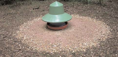
Fig 12 A . Mould in and around drinkers and feeders Considerable care has been taken to create a clean Area around the feeder.
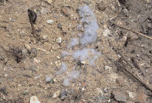
Fig 12 B. In spite of this care there is a considerable Mould growth immediately under the feeder
Mycotoxin inhibitors, often kaolin based, incorporated in food manufacture can dramatically reduce the risk.
Predator strike
Cause:
Mainly owls, most likely Tawny Owls. There are many other raptors, some of which are described as carrion feeders (e.g. Buzzards and even Red Kites) that have been seen to take live poults in and around the release pen.
Clinical signs:
Sudden death. Sometime birds have remarkably little external injury.
Treatment:
Nil
Prevention:
Provide adequate protection around shelters and plenty of hazards for making flight through the pens difficult, such as CD's suspended on nylon.
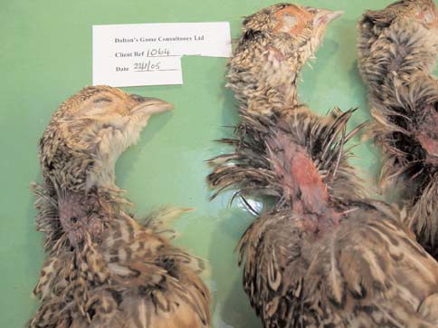
Figs 13A, B & C. Owl strike A. Neck lesions - external view
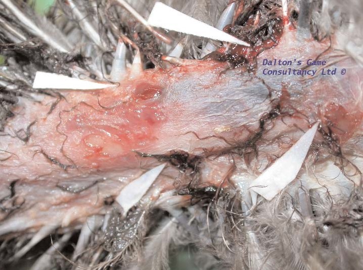
13B. Neck lesions dissected. White tabs indicate skin penetrations
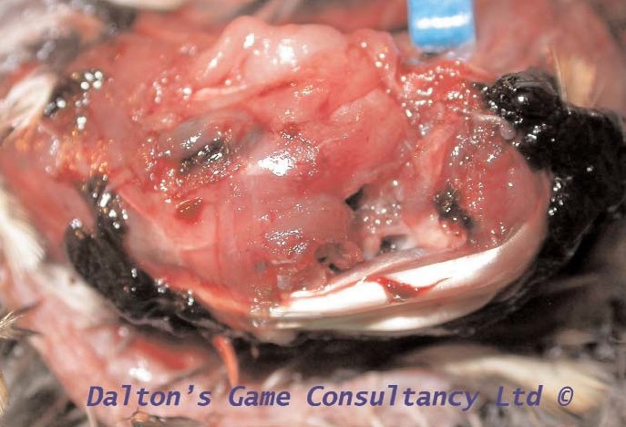
13C. Sub-cutaneous haemorrhages around head and neck
Trichomaniasis
Cause:
Trichomonads are single cell organisms that have become more and more important in causing disease in both pheasants and partridges. They are often found together with other parasites such as Hexamita (Spironucleus) and coccidiosis. Trichomonads are mainly found in the caecum but they may spill over into adjacent parts of the intestines and can also be found in the crop.
Clinical signs:
Presenting signs are similar to those caused by Hexamita, with weight loss, frothy yellow diarrhoea, weakness, dehydration and death. The diarrhoea is very similar to that seen in Hexamita and coccidiosis. It is not possible to make an accurate differentiation without examination.
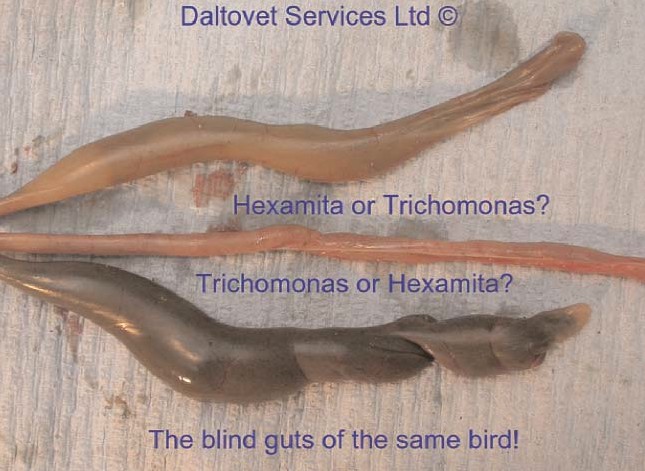
Fig 14 A Caecal (blind gut) contents variation in the same bird
The smaller (upper) blind gut is from a pheasant with Spironucleus and the larger (lower) had Trichomonas. Unfortunately, it is not often as clear cut as this, as is shown in the other picture. Trichomonas organisms are very small, being slightly larger than most bacteria. They can be detected under the microscope when moving. Therefore it is very important to bring in fresh samples as the organism starts to die soon after the bird has died and thus stop moving. Bringing in live, sick birds with signs typical for the flock problem will often aid diagnosis.
Post mortem:
Typically the blind gut (caecum) is grossly enlarged containing semi-liquid contents that vary in colour from pale yellow to khaki. Finding Trichomonas will not necessarily mean it is the cause of disease, as the organism can be found in the gut of perfectly healthy animals. The presence of very large numbers may be an indication of disruption of the normal caecal flora for other reasons.
Treatment:
The main aim is to provide supportive therapy with electrolytes and vitamins. The use of organic acids in water has proven to be advantageous. Oxytetracyclines and tiamulin do seem to help. In diseased birds this must be supplied in water, as the very first presenting sign is often loss of appetite, with associated rejection of feed, which may be scattered around the feeder. Oreganum extract (Herban®) is also believed to be of benefit. More important is prevention.
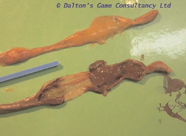
Fig 14 B. Caecal contents from birds with Hexamita - upper section, and Trichomonas - lower section, But they could look the other way round
Transmission occurs through bird-to-bird contact or through contact with contaminated feed, water or litter. Trichomonads cysts have not been identified and survive in small numbers in clinically normal birds. These can act as a source of infection for young birds in subsequent years. Wild birds can also introduce infection.
Prevention:
Prevention and control consist of good sanitation, husbandry and bio security measures.
Staphylococcosis
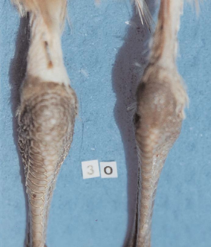
Figs 15 A, B & C. Staph Arthritis
A. Swelling around and below the joints
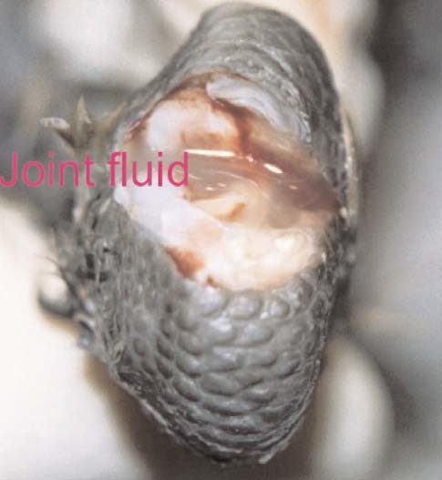
Fig 15 B. Opaque joint fluid (Staph Arthritis 0195A)
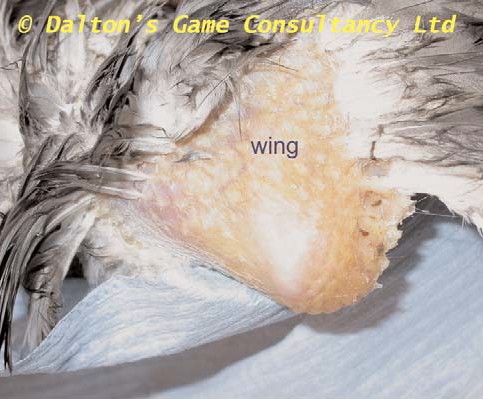
Fig 15 C Swollen wing joint with yellowing of the skin
Cause:
Infection, very often of the joints of the wings or legs, by Staphylococcus aureus. Rough handling at crating or damage to the skin by damaged wire or other metal work in the pens allows penetration of the skin by the bacterium. There is some evidence that poorly fitting bits and careless removal of them may also increase the risk.
Clinical signs:
Lameness, swelling around the joints. Discoloration of the skin.
Treatment:
Often antibiotics by injection needed in severe cases. Less badly affected birds will respond to antibiotic in water and/or feed.
Prevention:
Handle poults with considerable care. Make sure that wire is in good condition and don't allow scrap metal to build up in or near the pens. Take care when placing and removing bits.


